histological stains pdf
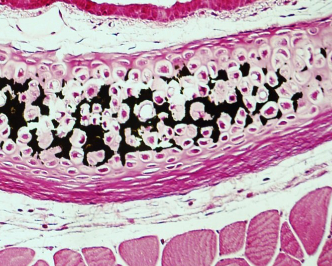
Fixation Any dichromate fixative Sections Thin paraffin sections. Black brown or golden stain.

Evaluation Of Quantitativity Of Histological Collagen Stains In Articular Cartilage Osteoarthritis And Cartilage
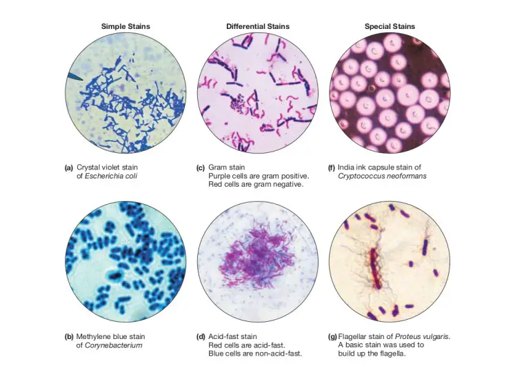
Acid-Fast Stain for Microorganisms 11.
. Staining techniques used were carmine silver nitrate Giemsa Trichrome Stains Gram Stain and Hematoxylin among others. Masson Trichrome - trichrome histology stains are formed from a mixture of three dyes. STAINS FOR MICROORGANISM STAINS COMPONENTS STAIN FUNGI 1.
CBSET histology specializes in the use of routine and customized staining techniques to ensure the most sophisticated analysis of tissue response to treatment for our partners. The techniques used can either be non-specific staining most of the cells in much the same way or specific selectively staining particular chemical groupings or molecules within cells or tissues. Red blood cells stain bright red - if cells have dark red staining in cytoplasm they are probably actively secreting acidophilic proteins.
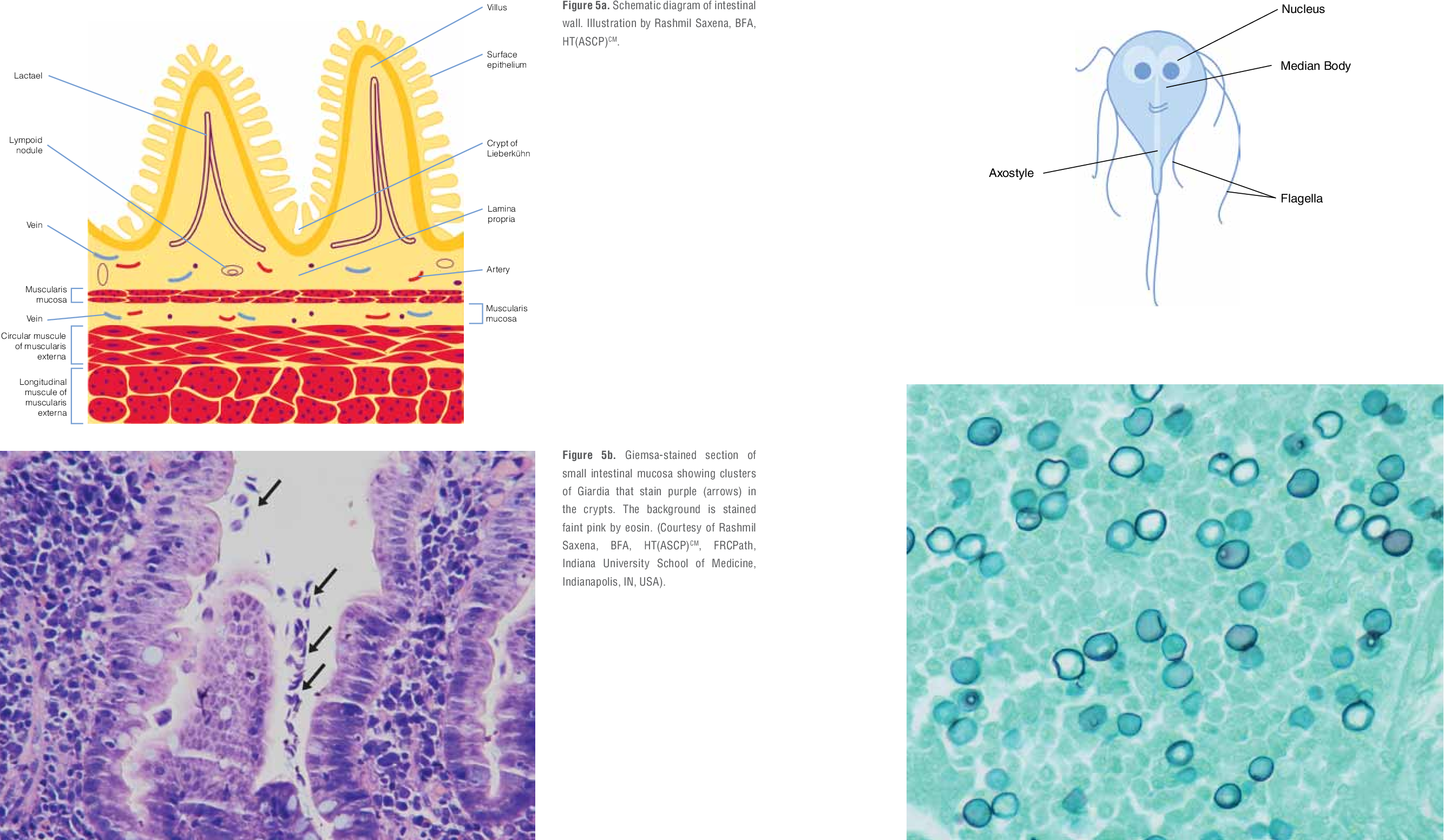
Warthin-Starry Stain for Microorganisms 9. Giemsa Usually used for staining blood and bonemarrow - smears. Stain with buffered Giemsa stain for 10 minutes dilute.
We glanced at the changes that have occurred in the histological staining process and advancement as well. Rarely used stains nuclei blue and cytoplasm - red. Histological stain available showing both cells and extracellular components.
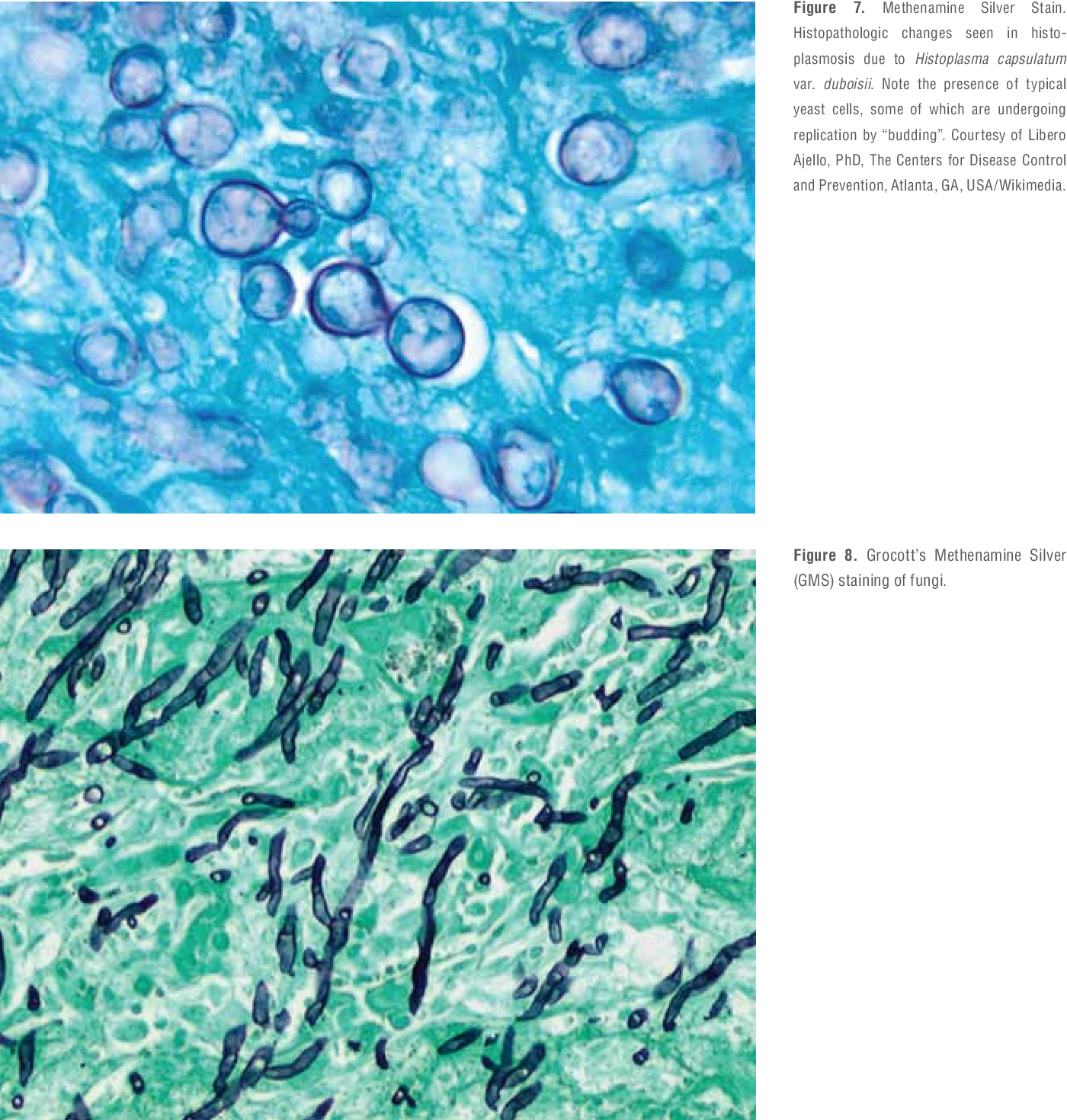
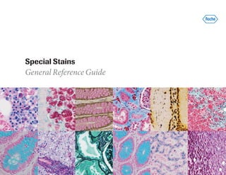
PAS FUNGI BLACK MUCIN TAUPE TO GRAY TISSUE GREEN FUNGI CELL WALL - MAGENTA Seen fairly well in HE but are demonstrated well with GMS and PAS. Fixation processing embedding sectioning and staining Titford 2009. Histological staining is a series of technique processes undertaken in the preparation of sample tissues by staining using histological stains to aid in the microscope study Anderson 2011.
For the pancreas glucagon secreting cells are stained pink and insulin secreting cells are stained blue. However many other special tinctorial stains are used on specific tissue or cellular components to aid diagnosis of toxicological or pathological changes. The most common stain applied for histological study is Haemotoxylin and Eosin.
Keywords artwork mere exposure taste preference histology. Histology stains are used to colour different structures within the cellsTissue processingBefore staining a slide the tissue has to be prepared and mounted onto a glass slideThe paraffin technique is the most common way to prepare a histological slide and follows the following stepsThe tissue sample is re-sectioned and fixed upon a slide. Peter Michalka Lucia Donárová Ústav patologickej anatómie LFUK a UN pracovisko Staré mesto Sasinkova 4 Bratislava Prof.
Different types of staining procedures used are given in the appendix. Histological staining acid basic histochemistry. The changes were made primarily to make histological staining easier faster cheaper and more accurate.
Aside from their utility in. The process of histological staining takes five key stages which involve. Review Methods A literature search on online medical databases for scholarly articles published from June 2011 to June.
Therefore we found a domain-independent exposure effect of images of histological stains to particular abstract paintings. Yeasts hyphae and spores Fungal cell walls are rich. After dichromate fixation the chromaffin granules stain a greenish-yellow colour with Romanowsky stains.
SILVER STAINS GROCOTT METHENAM INE SILVER NITRATE-GMS 2. Toluidine Blue Stain for Mast Cells Cancer Screening and Forensics 16. Certain of the stains use strong chemicals eg.
The purpose of this research was to assess past and current literature. This is a classic staining method for the adrenal. Nuclei are stained dark-blue to violet.
Histological sections have to be stained in some way to make the cells visible. Histological staining process and advancement as well as the likely reasons for these alterations. Useful in leukemia classification and elastic stains Figure 1B gauge a tumors degree of vascular invasion.
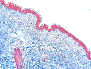
Gomoris Methenamine Silver GMS Stain for Microorganisms and Fungi 14. Other clinical applications for special stains cover a wide range of diseases. Mallory Trichrome - trichrome histology stains are formed from a mixture of three dyes - used on connective tissue to indicate collagen and reticular fibers.
This finding suggests that the taste for abstract art is altered by visual impressions that are presented outside of an artistic context. As a result histological staining is a multistep procedure that involves a variety of stains and other chemicals that may interact with other compounds found in tissues to change the results 3-6. Iron stains Figure 2A can indicate hemochromatosis or iron deficiency the Massons Trichrome stain Figure 2B demonstrates changes in collagen and.
The purpose of this research was to assess past and current literature reviews as well as case studies with the aim of informing ways in which histological stains have been improved in the modern age. Various staining procedures are applied from this hydrates stage. Various types of haemotoxylin formulations are used.
Carmine hematoxylin silver nitrate Giemsa trichome stain Gram stain and mauveine were among the first histological stains discovered in nature. The Hematoxylin and Eosin H. Gram Staining for Bacteria 7.
The expertise at CBSET can be utilized to develop the optimal processing sectioning and staining paradigm for a novel treatment device or material. Staining usually works by using a dye that stains some of the cells. PDF 98K Actions.
- uses acid fuchsine followed by a solution containing PTA orange G and aniline blue. The Bitesize Guide to Special Stains for Histology Contents 2. Staining techniques used were carmine silver nitrate Giemsa Trichrome Stains Gram Stain and Hematoxylin among others.
Use methanol fixed air-dried smears. CELL NUCLEI - stain darkly usually bluepurpleblack CYTOPLASM and CELL STRUCTURES - stain redpink.

Hematoxylin And Eosin Stain Stain Medical Videos H E Stain

Pdf Computational Histological Staining And Destaining Of Prostate Core Biopsy Rgb Images With Generative Adversarial Neural Networks Semantic Scholar
Types Of Staining Techniques Used In Microbiology Microbe Online
2

Pdf Introduction To Special Stains Semantic Scholar

An Intro To Routine And Special Staining In Histopathology

Pin On Histology And Anatomy
![]()
Histology Stains And Section Interpretation Kenhub
![]()
Histology Stains And Section Interpretation Kenhub

Bone Tissue With Osteoclast And Osteoblasts Stain Masson Goldner Trichrome Osteoclast Artsy Masson
Special Stains Department Of Pathology And Laboratory Medicine
Pdf Introduction To Special Stains Semantic Scholar

Pdf Comparison Of Special Stains For Keratin With Routine Hematoxylin And Eosin Stain Semantic Scholar
![]()
Histology Stains And Section Interpretation Kenhub
Special Stains Histology Research Core

Pin On What
Pdf Introduction To Special Stains Semantic Scholar

Pdf Dyes And Stains From Molecular Structure To Histological Application Semantic Scholar
Ventana Benchmark Special Stains General Reference Guide
0 Response to "histological stains pdf"
Post a Comment